A detailed diagnosis is necessary because it allows us to draw up an individual treatment plan for each and every patient. The visual material obtained helps to ascertain the condition of the teeth and present the course of treatment. We perform all of the diagnostics required for dental treatment at the clinic. This is extremely convenient and saves you time, and also makes it easier to maintain the consistency of the treatment and ensure the quality of the entire process.
Consultation and diagnostics prices
SPECIALIST CONSULTATION
50 €
ONLINE CONSULTATION
50 €
CONSULTATION WITH ORTHODONTIST E. BUČINSKIENĖ
80 €
ORTHODONTIST CONSULTATION
50 €
CONSULTATION WITH A PAEDIATRIC DENTIST
50 €
CONSULTATION FOR JAW JOINT DISORDERS
100 €
DENTAL X-RAY
10 €
PANORAMIC X-RAY
30 €
LATERAL CEPHALOGRAM
40 €
COMPUTED TOMOGRAPHY SCAN (3D)
100 €
DENTAL PACKAGE
10 €
NO-SHOW FEE
50 €
The prices shown are indicative. The doctor can only tell you the exact price after an individual consultation.
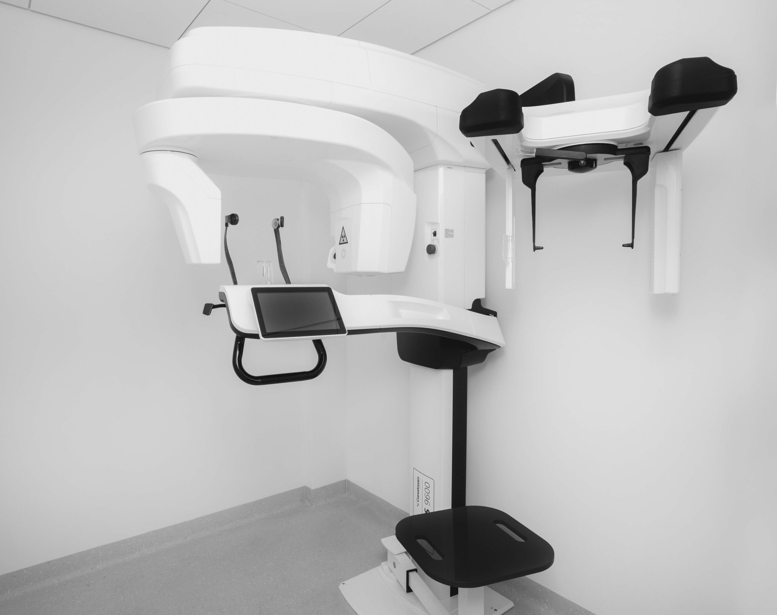
We have advanced tools that allow us to carry out detailed diagnostics.
This is the most accurate diagnostic method used in modern dentistry. This digital 3D image obtained shows a realistic view of the upper and lower jaws and the areas around them.
We use dental cone beam computed tomography when the 2D image of a regular dental X-ray is not enough – for example, before implant surgery.
All this allows us to draw up an individual treatment plan for implantation, endodontic treatment and other procedures. After everything is calculated and ready on the computer screen, all that needs to be done is to implement the carefully considered treatment plan in the dental chair.
According to the Radiation Protection Centre (RPC), the annual exposure of Lithuanian residents is determined by naturally occurring (radionuclides found in the Earth, ionising radiation from space, radionuclides in building materials, radon, a radioactive gas that escapes from the ground etc.) and medical (from diagnostic tests on patients using ionising radiation) radiation sources. Other factors determining exposure (activities with sources, radionuclides emitted into the environment from economic entities using sources and radionuclides present in food, drinking water, etc.) have less impact.
The average ionising radiation exposure worldwide is 2.9 millisieverts (mSv), and ranges from 1 mSv to 10 mSv per year, depending on the country (a millisievert is a calculated dose unit equivalent that reflects the biological effects of exposure). The second largest contribution to total exposure is exposure received during medical procedures, which is equal to 0.5 mSv per year on average.
In dentistry, the dose of ionising radiation received during a cone beam computed tomography examination is comparable to the natural radiation exposure accumulated over a period of 1-5 days.
In recent years, we have begun using many measures to reduce exposure. Computed tomography manufacturers have developed machines that provide excellent quality diagnostic images while exposing the person to a lower dose of radiation.
Therefore, you should not worry about the radiation you will get during the scan – precise and high-quality treatment that resolves your problem will bring much more benefits to you and your health than the harm from the scan itself.
These X-rays are also called “orthopantomograms”. They give a panoramic view of the upper and lower jaws and sinuses.
These are X-ray of one or more teeth that show the current situation in detail, for example, the size of the caries, the length and location of the roots, inflammation of the root canal and so on.
These are lateral X-rays that we use for orthodontic treatment.
Diagnostic dental models made of gypsum are intended for orthodontic treatment. Our clinic’s laboratory produces them according to the patient’s dental impressions. These models help to better assess the bite of the teeth, their position in the arches, proportions and so on. We look at the diagnostic models at the end of the treatment to evaluate the change – the treatment outcome.
This digital camera magnifies the image up to 100 times, so we can more accurately see the current situation and visually introduce you to it.
This device detects early changes in the teeth in relation to caries. Early diagnosis makes it possible to prevent more serious dental damage.
Treating teeth requires extreme care and precision, so we use image magnification equipment – a microscope. It makes our work easier and allows us to provide you with even more precise and high-quality treatment. At our clinic, we use the advanced Leica M320 dental microscope, which magnifies the image from 6.4 to 40 times.
Care and diligence are required when treating dental canals. The canals are not easily accessible and it is difficult to demonstrate their condition to the patient, so the patient often does not know what is happening during the treatment. The technologies that we use at our clinic allow us to show a full HD resolution image directly from your mouth and photograph the dental canals.
With the help of a microscope, we very carefully file the parts of enamel or dentine that are invisible to the naked eye and are damaged by caries, and then fill them. Dental restoration with the help of a microscope allows us to accurately restore the former shape of the tooth and guarantees a flawless aesthetic image after the procedure.
In microsurgical operations such as dental implants, gingivoplasty and so on, it is precision that determines how you will feel after the operation. Through constant monitoring and control of the operation with a microscope, we minimise the discomfort you feel after operations like these, and the operated area heals faster.
The microscope helps our clinical dental technologist not only to prepare crowns or dental bridges that fit perfectly, but also to maximally protect adjacent tissues. We rely on the microscope to check our calculations and evaluate the quality of the work done. The end result is a top quality crown, dental bridge or veneer, and your perfect smile.
Our dentist will decide what diagnostics you need. Depending on the case, the dentist may prescribe one or several tests.
USEFUL INFORMATION AND TIPS ON DENTAL HEALTH

Senior Dental Implantologist Marius Bučinskas attended a seminar in the USA

The clinic Dental Harmony took part in the charity event Christmas with Books

Awarded for educational activities

A training course Guided Implant Surgery & Aesthetics

A patient of ours shares her own personal experience about dental implant placement

A course on The Use of Micro-implants in Orthodontic Treatment